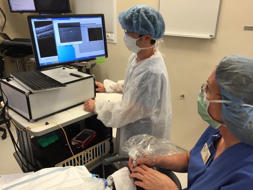
A team of engineers and physicians at Duke University has developed a new device that can capture high-quality images of retinas. It can produce high-resolution images of photoreceptor cells, or rods and cones.
Previous technology required the patient to sit still and concentrate for a few minutes, something children can't do very well. This lightweight handheld device fixes that problem.
"So the real significance of this device is it’s the first handheld device that can be used in infants and children who cannot sit still for long enough to be imaged using the conventional technique," said Dr. Joseph Izatt, professor of engineering and ophthalmology at Duke.
The project was the result of an ongoing collaboration between engineers and physicians at Duke. The project leaders – Izatt, Dr. Sina Farsiu and Dr. Cynthia Toth – have worked together in the past and they hope this new device will be a useful tool for researchers and clinicians alike.
"We hope that this will enable both better basic research pertaining to how the retina develops in infants and children, as well as potentially direct clinical applications in the diagnosis and treatment of blinding diseases that affect children just like they do in adults," Dr. Izatt said.
The technology still needs more testing, as well as a commercial manufacturer and FDA approval before it's distributed more broadly. That's a process that typically takes two or three years, according to Izatt. So far, the device has only been tested in a handful of adults and children.
Dr. Toth, a practicing clinician and retinal surgeon, will take over the continued testing.


